Aortic conditions represent some of the most demanding challenges in cardiovascular medicine, requiring a profound understanding of anatomy and surgical techniques. According to Dr. Vincenzo Giordano, these conditions, often involving aneurysms, dissections, or hereditary syndromes, can affect multiple parts of the aorta and necessitate personalized strategies. Recent innovations in imaging, surgical tools, and team-based care have greatly improved outcomes. High-volume centers with multidisciplinary teams are proving essential for managing these intricate cases. The integration of endovascular techniques, hybrid approaches, and data-driven planning continues to shift the treatment paradigm.
Complex Aortic Conditions
Aortic surgery focuses on treating serious disorders that affect the aorta, the body’s main artery. These conditions often involve aortic aneurysms, dissections, or congenital abnormalities that compromise blood flow and pose life-threatening risks if left untreated. Treatment decisions depend heavily on the location and severity of the disease. Thoracic aortic aneurysms, abdominal aneurysms, and dissections each require different approaches based on how they affect the vessel wall and surrounding structures.
Some patients may also have connective tissue disorders, adding more complexity to surgical planning. In such cases, genetic counseling and family screening may also be advised. In some cases, surgeons must address multiple sections of the aorta during a single procedure. Conditions like Marfan syndrome or Loeys-Dietz syndrome frequently involve the entire aortic tree, requiring a staged or hybrid approach to care tailored to the patient’s anatomy and disease progression.
Surgical Challenges and Risk Factors
Treating complex aortic diseases involves navigating numerous anatomical and physiological hurdles. The aorta’s proximity to vital organs and its branching structure increase the risk of complications such as stroke, spinal cord injury, or organ ischemia during surgical intervention. The delicate balance between aggressive intervention and preserving organ function makes each case unique.
Each patient presents a unique clinical picture, which can dramatically influence surgical planning. Age, underlying health conditions, prior surgeries, and the presence of inflammatory or connective tissue disorders all affect how the body responds to surgery and recovery. Surgeons must weigh these variables carefully to reduce risk and improve outcomes. In re-operative cases, scar tissue and altered anatomy compound the challenge.
Emergencies like aortic rupture or acute dissection add urgency and complexity, especially when patients arrive unstable or with limited history available. Decisions must be made swiftly, and the margin for error is minimal.
Changes in Surgical Methods and Technologies
Advances in surgical technique have broadened treatments for complex aortic conditions. While traditional open surgery remains essential for certain cases, endovascular repair has become more prevalent, offering reduced recovery times and lower immediate surgical risk. These minimally invasive approaches are now being used in anatomically difficult areas once considered inoperable.
Hybrid procedures combine the strengths of open and minimally invasive approaches. In cases involving the aortic arch or thoracoabdominal segments, surgeons may use custom stent grafts and debranching techniques to preserve blood flow to critical arteries while minimizing trauma. Some institutions have developed dedicated hybrid operating rooms to facilitate these interventions.
Modern imaging and intraoperative guidance tools now play a pivotal role in planning and execution. High-resolution CT scans, 3D reconstructions, and real-time navigation systems allow for greater precision, enhancing the safety and effectiveness of increasingly complex procedures.
Enhancing Surgical Precision
Aortic surgery has been transformed by innovations that improve accuracy and ease invasiveness. Graft materials are now designed for better durability and flexibility, allowing surgeons to tailor repairs to the patient’s anatomy with greater confidence. Custom-manufactured stent grafts have also expanded the feasibility of endovascular repair in anatomically challenging cases. These materials are often engineered to reduce inflammatory response and enhance long-term integration into the vessel wall.
Techniques such as 3D anatomical modeling and pre-operative simulation have shortened learning curves and improved surgical planning. In select centers, robotic-assisted systems are being explored to provide enhanced dexterity during intricate aortic reconstructions. These tools help refine incision placement, device deployment, and vascular control, especially in confined or high-risk areas.
Minimally invasive techniques continue to gain ground in treating high-risk patients who may not tolerate open procedures. By accessing the aorta through smaller incisions or peripheral arteries, surgeons can significantly reduce trauma and shorten recovery without compromising long-term outcomes. This is particularly beneficial for elderly patients or those with multiple comorbidities.
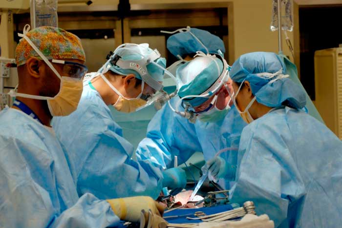
Role of Specialized Teams and High-Volume Centers
Outcomes in aortic surgery are closely tied to the experience and coordination of the medical team. High-volume centers with dedicated vascular and cardiothoracic units tend to achieve better results, owing to familiarity with rare complications and the availability of advanced support services. These institutions often have protocols in place for rapid escalation of care and access to specialized equipment.
Team-based care is paramount, especially during hybrid or staged procedures. Collaboration between surgeons, anesthesiologists, radiologists, and critical care specialists ensures that every phase of treatment—from diagnosis to recovery—is guided by shared expertise. Institutions that invest in simulation labs, ongoing education, and multidisciplinary case reviews are better equipped to handle challenges in aortic care.